





Corneal Conditions

The cornea is the clear, dome-shaped surface at the front of your eye. It protects the eye and helps focus light so you can see clearly. The cornea has several layers, and problems can affect any of them.
Corneal problems can cause pain, redness, sensitivity to light, blurry vision, or loss of vision. Some conditions are caused by an injury or infection. Others develop because of inherited conditions, aging, inflammation, or changes in the cells that keep the cornea healthy.
At Eye Associates, we diagnose and treat many conditions of the cornea and the surface of the eye.
Corneal Abrasion
A corneal abrasion is a scratch on the surface of the cornea. It can happen from a fingernail, piece of paper, contact lens, foreign object, or other injury.
The cornea has many nerves, so even a small scratch can be very painful. Symptoms may include:
- Eye pain or a feeling that something is in your eye
- Tearing
- Redness
- Sensitivity to light
- Blurry vision
Most small abrasions heal within a few days. Treatment depends on the cause and severity of the injury. Your eye doctor may prescribe medication, including an antibiotic when appropriate, to help prevent infection.
Do not rub your eye or try to remove an object that is stuck in the eye. A deeper injury or an abrasion that does not heal normally should be examined by an eye doctor.
In some people, an old abrasion can lead to recurrent corneal erosion, in which the surface layer of the cornea repeatedly comes loose.
Corneal Ulcer
A corneal ulcer is an open sore on the cornea. It is a serious condition because an ulcer can cause scarring and permanent vision loss if it is not treated quickly.
Corneal ulcers are often caused by an infection. People who wear contact lenses have a higher risk, especially if they sleep in their lenses, do not clean them properly, or use them longer than recommended.
Symptoms can include: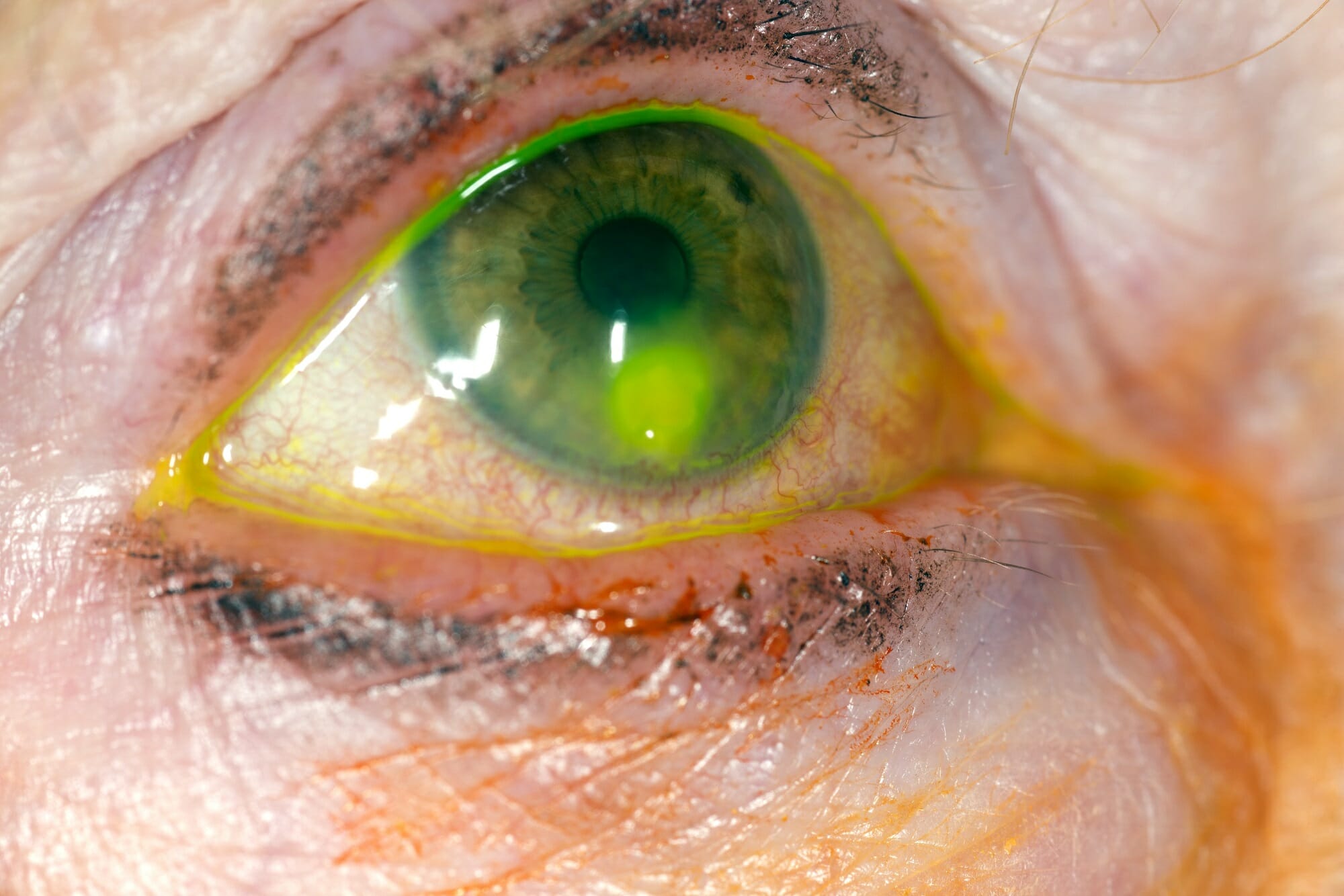
- Eye pain
- Redness
- Tearing
- Sensitivity to light
- Blurry or decreased vision
- A white or cloudy spot on the cornea
A corneal ulcer needs prompt medical attention. Treatment depends on the cause and may include antibiotic, antiviral, or antifungal medication.
If you wear contact lenses and develop eye pain, redness, or decreased vision, remove your contact lenses and contact an eye doctor promptly.
Recurrent Corneal Erosion
Recurrent corneal erosion happens when the outer layer of the cornea, called the epithelium, does not stay firmly attached to the layer underneath it.
It can occur after a previous corneal scratch or with certain corneal conditions, including epithelial basement membrane dystrophy (also called map-dot-fingerprint dystrophy).
A common symptom is sudden eye pain when you first open your eyes in the morning. Other symptoms may include:
- A feeling that something is stuck in the eye
- Tearing
- Blurry vision
- Sensitivity to light
Treatment depends on how often the erosions occur and how severe they are. Lubricating eye drops and ointments are often used. Your doctor may recommend other treatments if the problem continues.
Map-Dot-Fingerprint Dystrophy
Map-dot-fingerprint dystrophy, also called epithelial basement membrane dystrophy (EBMD), is a common condition affecting the outer layer of the cornea.
The surface of the cornea can develop patterns that look like maps, dots, or fingerprints. In many people, this causes few or no symptoms. In others, the surface does not attach normally and can lead to recurrent corneal erosions.
Symptoms may include:
- Blurry or changing vision
- Eye irritation
- A feeling that something is in the eye
- Pain, especially when waking up
- Sensitivity to light
Treatment may include lubricating eye drops or ointment. If symptoms are severe or continue despite medical treatment, procedures such as superficial keratectomy or phototherapeutic keratectomy (PTK) may be considered.
Keratoconus
Keratoconus is a condition in which the cornea becomes thinner and gradually bulges outward into a cone-like shape.
Because the cornea helps focus light, changes in its shape can cause blurry or distorted vision. Keratoconus often affects both eyes, although one eye may be worse than the other.
Signs and symptoms can include: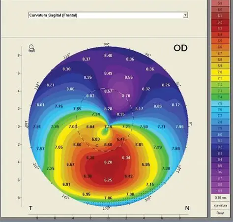
- Blurry or distorted vision
- Frequent changes in glasses prescriptions
- Difficulty seeing clearly at night
- Increased sensitivity to light
- Difficulty wearing contact lenses
Treatment depends on how advanced the condition is. Glasses or contact lenses can often improve vision. Special rigid or scleral contact lenses may be helpful when the cornea becomes more irregular.
A treatment called corneal cross-linking can strengthen the cornea and may slow or stop the progression of keratoconus in appropriate patients.
If keratoconus becomes advanced and causes severe scarring or vision problems that cannot be corrected with contact lenses, a corneal transplant may be needed.
Avoid rubbing your eyes, especially if you have keratoconus or are at risk for it. Your eye doctor can recommend treatment for allergies or other conditions that cause eye itching.
Fuchs' Endothelial Corneal Dystrophy
Fuchs' endothelial corneal dystrophy is a condition that affects the innermost layer of the cornea, called the endothelium.
Endothelial cells help keep the cornea clear by pumping excess fluid out of it. These cells normally do not replace themselves very well. When too many cells are lost or stop working properly, fluid can build up in the cornea. This causes corneal swelling, also called edema, and can make vision cloudy.
Symptoms may include:
- Blurry vision, often worse in the morning
- Glare or halos around lights
- Difficulty seeing at night
- Sensitivity to light
- Cloudy vision
- In more advanced cases, eye pain
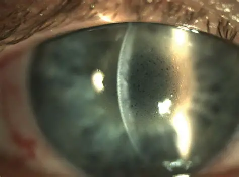
Treatment depends on the severity of the disease. Mild cases may be treated with special eye drops or ointments that help reduce corneal swelling. When the endothelial cells are no longer able to keep the cornea clear, an endothelial corneal transplant may be recommended.
Endothelial Corneal Transplant
Not every corneal transplant requires replacing the entire cornea.
The endothelium is the thin layer of cells on the back surface of the cornea. These cells act like tiny pumps. They remove extra fluid from the cornea and help keep it clear.
When the endothelium becomes severely damaged, the cornea can swell and vision can become cloudy. This can happen with Fuchs' dystrophy, after certain types of eye surgery, or from other causes of endothelial damage.
An endothelial keratoplasty replaces the damaged inner layer with healthy donor tissue. Because only the damaged inner layers are replaced, most of the patient's own cornea remains in place.
Two common types are DMEK and DSAEK.
DMEK
DMEK (Descemet membrane endothelial keratoplasty) replaces the damaged endothelial cells and their supporting layer, called Descemet's membrane.
The donor tissue is extremely thin. It is placed inside the eye and positioned against the back of the patient's cornea. An air or gas bubble is used to hold the tissue in place while it attaches.
DMEK is commonly used for conditions such as Fuchs' endothelial corneal dystrophy and other diseases that cause endothelial failure.
DSAEK
DSAEK (Descemet stripping automated endothelial keratoplasty) also replaces the damaged endothelial cells, but the donor tissue includes the endothelium, Descemet's membrane, and a very thin layer of the back part of the cornea.
Like DMEK, the donor tissue is placed inside the eye and supported with an air or gas bubble while it heals.
What is the difference between DMEK and DSAEK?
Both procedures replace the part of the cornea that is not working properly. The main difference is the amount of donor tissue used.
- DMEK: endothelial cells + Descemet's membrane
- DSAEK: endothelial cells + Descemet's membrane + a thin layer of corneal tissue
Your surgeon will determine which procedure is most appropriate based on the condition of your cornea and your individual needs.
Endothelial transplantation does not correct every type of corneal problem. It is designed for diseases caused by a poorly functioning endothelium. Conditions involving major corneal scarring or severe corneal thinning may require a different type of corneal transplant.
Pterygium
A pterygium is a non-cancerous growth of the conjunctiva, the tissue covering the white part of the eye. It can grow onto the cornea.
Pterygia are often associated with long-term exposure to sunlight, wind, dust, and other environmental irritation.
A pterygium may cause: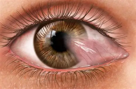
- Redness
- Irritation or a gritty feeling
- Dryness
- Changes in vision if it grows onto the cornea
Artificial tears and protection from ultraviolet (UV) light may help with symptoms. If a pterygium continues to grow, causes significant irritation, changes the shape of the cornea, or affects vision, surgery may be recommended.
Ocular Herpes
Ocular herpes is an infection of the eye caused by herpes simplex virus, the same family of viruses that can cause cold sores.
Herpes can affect different parts of the eye. When it affects the cornea, it can cause inflammation and, in some cases, scarring.
Symptoms may include:
- Eye redness
- Pain or irritation
- Tearing
- Sensitivity to light
- Blurry vision
Treatment depends on which part of the eye is affected and how severe the infection is. Antiviral medication may be prescribed as eye drops, ointment, or pills.
Because herpes infections can return, follow-up with an eye doctor is important.
Allergic Conjunctivitis
Allergic conjunctivitis affects the conjunctiva, the thin tissue that covers the white part of the eye and lines the eyelids. It is caused by an allergic reaction.
Common symptoms include:
- Itchy eyes
- Redness
- Watery eyes
- Swelling
- Burning or irritation
Allergic conjunctivitis usually does not damage the cornea, but severe eye rubbing can cause problems with the surface of the eye and may worsen conditions such as keratoconus.
Treatment may include avoiding allergens, artificial tears, antihistamine or other allergy eye drops, and other medicines when needed.
Conjunctivitis (Pink Eye)
Conjunctivitis, commonly called pink eye, is inflammation or infection of the conjunctiva. It is different from a disease of the cornea.
Viruses are a common cause of pink eye. Bacteria and allergies can also cause conjunctivitis.
Symptoms may include:
- Red or pink eyes
- Watery or sticky discharge
- Irritation or burning
- Itching, especially with allergies
Many viral cases improve without specific treatment. Antibiotic eye drops may be prescribed for certain bacterial infections.
Because several different conditions can cause a red eye, an eye examination may be needed to determine the cause.
Corneal Transplantation
If the cornea is severely damaged or cloudy and other treatments cannot restore useful vision, a corneal transplant may be recommended.
There are different types of corneal transplantation. The type depends on which layers of the cornea are damaged.
A full-thickness corneal transplant, called penetrating keratoplasty (PK), replaces all layers of the central cornea.
A partial-thickness transplant replaces only the damaged layers. Examples include endothelial procedures such as DMEK and DSAEK.
Using a partial-thickness transplant when appropriate allows the surgeon to preserve more of the patient's healthy cornea.
Corneal transplant surgery is performed using donated corneal tissue. Recovery depends on the type of transplant and the health of the eye.
After surgery, patients use prescription eye drops and have regular follow-up visits. It is important to avoid rubbing or pressing on the eye and to follow all postoperative instructions.
Signs of Possible Corneal Transplant Rejection
Corneal transplant rejection can sometimes be treated successfully when it is recognized early.
Call your eye doctor promptly if you have a transplanted cornea and develop:
- New or increasing eye redness
- Eye pain
- Increased sensitivity to light
- Cloudy or hazy vision
- A noticeable decrease in vision
Do not wait for these symptoms to go away on their own.
When Should I See an Eye Doctor?
Seek prompt medical attention for:
- Sudden changes or loss of vision
- Significant eye pain
- A red, painful eye
- New sensitivity to light
- A white spot on the cornea
- Eye pain or decreased vision while wearing contact lenses
- An eye injury, especially from a sharp object or chemical
- Symptoms that are getting worse instead of better
Early diagnosis and treatment can help protect the cornea and preserve vision.


If you would like more information about Corneal Conditions or to schedule an appointment, feel free to fill out our convenient contact form or call us directly at 215-997-2015.